[Perspectives of Traditional Chinese and Western Medicine]Reporting errors affect the direction of treatment, imaging examinations for cancer patients, and communication between doctors is the key
[ad_1]
Text ◆ Su Ziqian (clinical oncology specialist/TCM physician)
After all, for cancer patients, whether for diagnosis or treatment follow-up, they often need to take different images such as computer scan (CT), magnetic resonance imaging (MRI), and positron emission tomography scan (PET CT).
In fact, are the photos the same wherever you go? ? Is price the only consideration?
(Hong Kong News) Medical imaging is becoming more and more developed and more common. Not only patients sometimes need to take different images, some healthy people also undergo physical examinations and simple medical imaging tests such as lung X-rays or abdominal ultrasounds.
First, let’s briefly talk about the functions of different medical images. The simplest is to take X-rays of the lungs. Generally, X-rays of the lungs have great limitations in diagnosing lung cancer or clearly seeing the stage of the disease. The reason is that the resolution of X-rays is too low.
However, X-rays of the lungs are still useful. They are generally useful for follow-up treatment of diagnosed cancer (especially lung cancer), or for preliminary examination of patients who often cough and asthma.
X-rays are cheap and have low radiation resolution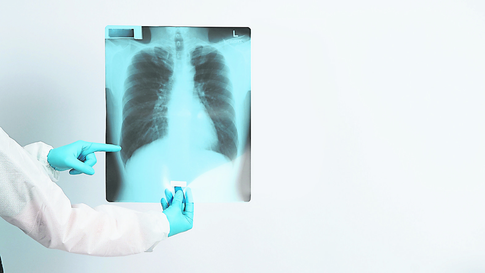
The advantage of X-ray is that it is cheap and has low radiation. However, due to the low resolution of lung X-ray, it is basically impossible to see early lung cancer. Therefore, even if smokers are currently screened for lung cancer, international guidelines recommend the use of computer sketches of the chest and lungs rather than the use of X-rays. So simply put, X-rays cannot see many things.
In addition, computer sketch CT is a relatively high-energy X-ray and multi-slice image. It is generally used for examination of the lungs and abdomen. For example, it can be used to check whether there are tumors in the lungs and liver, ascites and pulmonary fluid. It also has a great prompting effect. Sometimes it is also used on the bones of the limbs to see if the bones have been eroded by tumors. It also has a certain effect. When taking a computer sketch, you can choose to take it as it is or apply a developer (the scientific name is called enhanced computer sketch). In many cases, tumors have many blood vessels, so after applying the developer, some tumors that are similar to normal cells on computer scan images will appear smaller. Easy to show.
So when do you need to apply developer? Generally, liver tumors, whether they are primary liver cancer or have spread to the liver, also require contrast agent examination. In addition, if it is lung cancer that is planned to be operated on, the patient needs to clearly see the relationship between blood vessels and lung tumors, so it is generally recommended to use a contrast agent. But please note that if the kidney function is not good, you should be careful when using the developer.
PET CT shows whether the tumor is active or not
In addition, cancer patients are more likely to undergo positron emission scans (PET CT). In fact, most cancer diagnosis and treatment follow-up require PET CT if possible. In many cases, CT alone is much better. Compared with ordinary CT, PET CT will inject some radioactive drops into the patient, so the activity of different tumors can be seen.
To put it simply, if CT alone can only see the size and shape of the tumor, PET CT can also see whether the tumor is active or not in addition to the size and shape of the tumor. Because sometimes some tumors will leave some scars after treatment, whether with drugs or electrotherapy. If CT is used alone, sometimes it is impossible to tell whether the tumor is still active or whether it is basically a post-treatment change of the tumor.
That’s why most current cancer treatment follow-up generally requires regular PET CT. Pay attention to the medicine mentioned. The most common one is a special sugar FDG, but there are other medicines such as PSMA, which is specifically used to treat prostate cancer. (In fact, cancer does not suck sugar. The fallacy that cancer sucks sugar has been wrong for decades. Readers can refer to a long article to explain this.) But of course the disadvantages of PET CT are that it is more expensive and the radiation dose is generally higher than that of CT.
MRI is very helpful to see changes in bone marrow
In addition, magnetic resonance MRI is an examination method without radiation. It is generally used for imaging examinations of the brain and spine, especially for gynecological and prostate examinations. Sometimes magnetic resonance imaging can also be of great help to see changes in bone marrow. Magnetic resonance imaging can also use a developer like CT (but the developer is different).
So, when cancer patients undergo various imaging examinations, is price the only consideration?
A few months ago, a patient with stage III lung cancer came for a follow-up consultation. The patient has received chemotherapy and immunotherapy and has completed six weeks of electrotherapy. He is receiving maintenance injections of immunotherapy for one year, and is also taking traditional Chinese medicine for anti-cancer treatment. About three months after the electrotherapy, a positron scan was performed to see the reaction after the treatment.
After a preliminary report written by a radiologist, it was found that the area where the electrotherapy was performed looked like the area where the tumor had undergone electrotherapy was very bright and much larger. So has the disease gone out of control after treatment failed? Later, the author discussed the case with the radiologist. After careful review and analysis over time, I found that the patient’s cough and asthma were better than before.
Specialists need clinical data
Therefore, the subsequent report concluded that those areas where the lungs were damaged were the side effects of pneumonia three months after normal electrotherapy. This is a normal reaction of the body after tumor necrosis, and it does not mean that the disease has become worse. Later, I told the patient that he didn’t need to worry about continuing the original treatment. Three months later, he followed up with a regular X-ray and found that the spots where he was suffering from pain gradually subsided on their own, just as expected.
It can be seen that for a good imaging report, the radiologist needs to have sufficient clinical information from the clinician. On the other hand, follow-up requires two doctors to be able to communicate directly, otherwise the slightest difference can lead to a huge mistake! Think about it, if an unqualified report mistakenly believes that the radiotherapy was spent on lung cancer, then the patient may have received more chemotherapy that was not needed, completely affecting the treatment direction of the disease.
Many patients think that,
In fact, the photos are the same everywhere.
Yes, although there are old and new instruments,
But the most important thing is not the instrument.
A lot of CT, MRI,
Or even changes in PET CT,
In fact, in many cases, absolute conclusions cannot be given.
When radiologists generally consider how to report
The type of tumor of the patient should also be considered.
Changes after previous treatments,
Only then can the conclusion of the report be written.
Sometimes a good report will write about what is most likely to be, but there are also less likely to be another scenario. Because many changes seen in imaging are not medically absolute. If the doctor does not consider the patient’s condition and compare it with previous old films, it is actually difficult to make a good conclusion.
Especially now that there are more and more cancer treatments and drugs, many tumors also have some special imaging changes after treatment. Therefore, radiologists and oncologists actually communicate closely before issuing a report. I myself often receive calls from radiologists asking for more information before issuing a report. When I see some areas that are unclear in the report or CT scan, I often call the radiologist to inquire and discuss them again.
Therefore, in fact, every time a patient undergoes an MRI, communication and cooperation between the attending physician who is responsible for writing a referral to the imaging examination and the radiologist who is responsible for the report are also very important.
Treatment follow-up reports should be compared
People compare with others. As the saying goes, there is no harm if there is no comparison. But for imaging reports, it really doesn’t hurt if there is comparison.
For cancer patients, PET CT is often used as a follow-up treatment before and after treatment to see whether the condition is getting better, worse, or stable due to drug treatment, etc. The best report is with previous imaging as a check. Sometimes the report will have a list, detailing, for example, six places where tumors have spread in the past, how the size and activity of each place has changed after treatment.
There is usually a detailed list for comparison. In addition, whether any new diseases have emerged will usually be written clearly. Therefore, there must be comparison in the treatment follow-up reports of cancer patients! Otherwise, it will be really difficult for clinicians to follow up.
Depending on the disease Re-evaluate treatment directions
It is important to know that no matter what imaging examination is done, the ultimate goal is to see the changes in the disease, so that the oncologist can know whether it is necessary to change the treatment, maintain the treatment, or even reduce and stop the treatment.
There is no comparative report, which is a bit like taking photos in vain, and it is of little benefit to clinicians and patients.
So if we took photos in the same place in the past, will we have to take photos in the same place in the future? In fact, after CT, MRI and PET CT are generally performed in Hong Kong, a DVD or a USB drive containing the DiCOM files of the image is usually included in the report. These imaging data are actually used internationally, so, If the second radiograph is not taken at the same place, as long as you take the report and the CD or finger with you when you go to another place to take the radiograph, the general radiologist can load the images into his or her computer system for comparison.
Misjudgment wastes money
Sometimes some patients may not have had these imaging tests done in Hong Kong, and some radiology reports do seem to be inferior to Hong Kong standards. Patients will put DVDs or USBs in the clinic, hoping that clinicians can help them make comparisons. But in fact, it is another specialist who is responsible for comparing imaging and generating conclusions, that is, a radiologist.
This is a very professional and complex subject. Whether it is in terms of the patient’s interests or liability issues, it is difficult to draw a responsible conclusion based solely on comparisons made by clinicians. Moreover, it is generally difficult for Hong Kong doctors to communicate directly with doctors from other places who write reports. In addition, doctors from other places do not necessarily have Hong Kong radiology specialist qualifications.
In some cases, for the sake of safety, such patients need to take another test when they return to Hong Kong.
I often tell patients that every time before deciding to take any scan, clinicians will actually consider whether it is necessary, which method is better, and when to take it. If in some cases it is necessary to use PET and CT for comparison, but in the end the patient only uses CT, the final conclusion cannot be drawn and the patient will have to be re-examined. On the one hand, it is a waste of money, and on the other hand, it is exposed to unnecessary radiation. Try to avoid this situation from happening.
Consult your doctor before making a decision
For some more complex cases, doctors sometimes take the patient’s films and reports into meetings with other doctors and radiologists to re-examine the direction of treatment. This situation is generally more common before surgical planning or electrotherapy planning.
No matter what sketch is taken by the patient, the attending physician who wrote the referral paper for the patient must be consulted before making a decision.
[ad_2]
Source link
![[Love Wants Sexual Happiness Series 358]Find the culprit and overcome psychogenic erectile dysfunction. Don’t let pressure affect your sexual happiness.](https://chinathenews.com/wp-content/uploads/2024/04/171111-780x420.jpg)

![[Wanqingyi Care]My health, my rights, customized medical methods in the last stage of life](https://chinathenews.com/wp-content/uploads/2024/04/ZZ1-100-780x420.jpg)
![[Kidney Transplantation Special Topic]The survival rate of transplanted kidneys is high without dialysis treatment three times a week](https://chinathenews.com/wp-content/uploads/2024/04/1311-780x420.jpg)