
[Love Wants Sex Series 312]Mammography plus ultrasound is more accurate in detecting breast cancer
[ad_1]
Transcript: Liang Yingxiu
(Kuala Lumpur News) There are many types of breast examinations, which one is suitable for every woman?
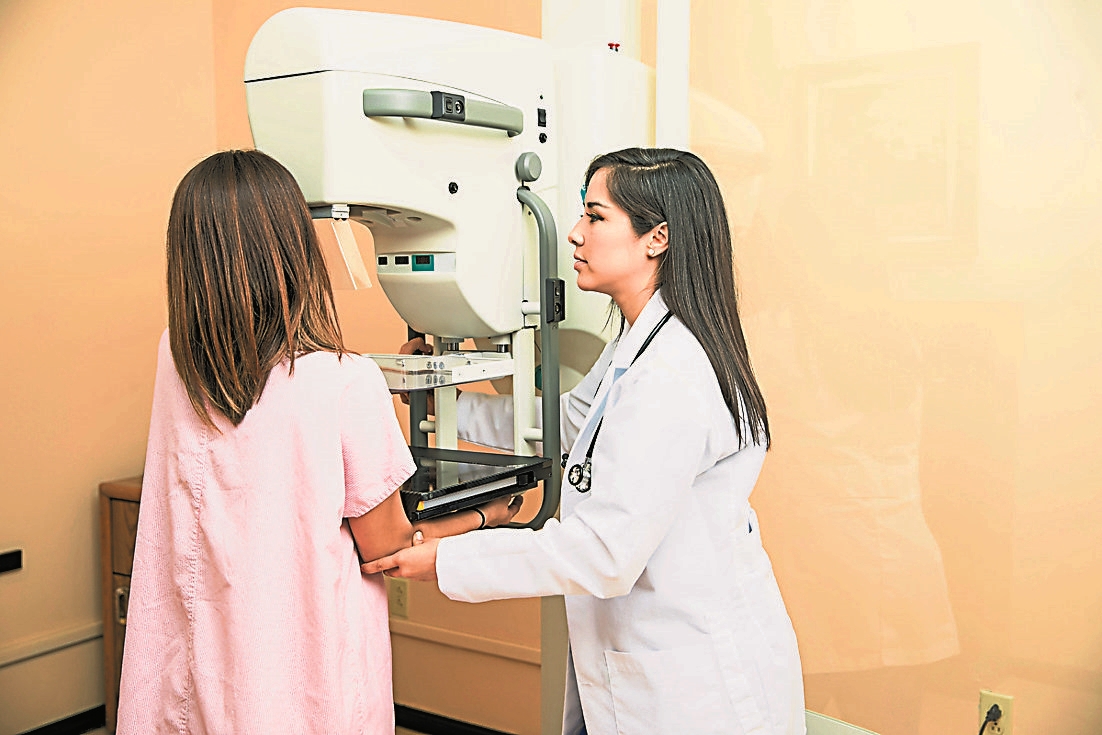
Breast surgery consultant Dr. Chen Yifei pointed out that there are many factors for us to consider. The first breast examination method that is generally seen on the market is the common mammography or breast Xray, which is called Mammogram in English, which is also commonly known as ” The second is B-ultrasound, which is an ultrasound examination; the third is nuclear magnetic resonance (MRI).
Mammography is sensitive to calcifications
Many people get confused about when to have a mammogram; or when to have an ultrasound. Is doing one enough? Or do two? Which one is more useful or effective and can be seen more clearly? If you do both, you will worry about more radiation.
For all these questions, she said, in fact, these two kinds of examinations are different in observing breast tissue. Formally speaking, a complete breast examination is a combination of mammography first and then ultrasound examination, because doing both mammography and ultrasound examination will give greater accuracy!
“It can tell whether the nature of a tumor is good or bad.”
She explained that, first of all, mammography is more suitable for transillumination imaging of breast soft tissue. It is particularly sensitive to calcifications. Sometimes doctors will tell patients to look at these white spots, which are calcifications, which are called micro calcifications in English.
“Because most early breast cancers, before granules have formed, it may appear as calcifications. So, this is the reason for detection by mammography. However, not all early breast cancers may appear as calcifications , Therefore, at this time, it is necessary to combine ultrasound examinations to accurately diagnose the existence of cancer.”
She pointed out that secondly, mammography examination is very affected by breast density, which we call breast tentative. Because for young women, her glands are very dense and fibrous tissue is abundant, so the dense shadow of the entire breast layer shown by photography may not have a sense of layering, and it is very difficult to find the existence of calcification points; Under the sound wave examination, its clarity will be relatively high, so it can effectively show the relationship between the internal structure of the breast cancer tumor and the internal tissues around the tumor, and sometimes even the size, shape and edge of the lesion may be seen .
“At the current medical level, ultrasound examination can see tumors of 0.3 to 0.5 centimeters, which can be said to be a simple, convenient and non-invasive examination.”
She said that from this statement, I hope that everyone can easily distinguish the difference between mammography and ultrasound. In fact, each has its own advantages and disadvantages, so in many cases we have to cooperate with each other to obtain higher accuracy.
I heard that the examination will be very painful. Are you keen?
Many women are not very keen on mammography examination, because they often hear that the examination is painful and very uncomfortable, so they generally prefer to do ultrasound examination.
Is it really painful? She said, first of all, you need to understand that the breasts will definitely be compressed during the photographic examination, so do not perform the examination when the breasts are the softest, that is, do not perform such examinations one week before the menstrual period and during the menstruation. It is best to do it one week after the menstrual period; even if you have breast pain or it hurts when you press it gently, you can take some painkillers orally to relieve the pain before proceeding.
“Before mammography, you are usually warned not to use deodorants, antiperspirants, foundations, creams, face creams or perfumes on the underarms and around the breasts because these items contain metal particles that may damage the breasts. It is different in photography, so it will affect the accuracy.”
She stressed that the inspection only takes a few minutes to complete. During the whole process, it is very important to communicate with the technician who performed your breast examination. If you feel very uncomfortable, you can tell her to slow down and squeeze the breast slowly so that you will not feel pain.
Mammography Safety
Only 10% report abnormal
The dose of mammography examination is very low and safe, that is, it will not have a great impact on us. After the mammography examination report, about 10% of women will have abnormal results, that is, abnormal , need further inspection.
In common cases, tumors can be seen on some planes, but it is still unclear whether it is good or bad, benign or malignant?
For example, Chen Yifei said that if 1,000 women undergo mammography examinations, about 100 of them will be recalled for further examinations. Among them, they will undergo ultrasound examinations, and among the 100 recalled women, 10 will need to undergo biopsy , according to the percentage rate, about 3% of people will be diagnosed with breast cancer.
Breast calcification is common and there is no need to be nervous
“When do you usually call a patient back for further examination? If the doctor sees calcifications in milk ducts or tissue, suspicious calcifications, lumps, distorted tissue, or dense shadows in the breast that were not there at all last time. This phenomenon; or if the shadow is not found in the previous image inspection, it may be a new discovery, and further inspection may be needed.”
But not all calcifications are dangerous? “Calcification may be caused by cell secretion, cell fragments, inflammation or trauma; or some patients who have undergone breast implant surgery or used breast enhancement products may have calcification.
This calcification point has nothing to do with the calcium tablets we take. Generally, there are two types of calcifications: the first is micro calcifications, which are very tiny and irregular calcifications, which are usually related to breast cancer; the other is macro calcifications, which are relatively rough and relatively large calcifications. These are usually benign lesions that may have become calcified due to aging, trauma, or fibrous cysts.
“Therefore, when you find calcification after examination, don’t be nervous, because breast calcification is actually very common. Many women have one or more calcifications, and most of the calcifications are benign lesions. If there are malignant phenomena , the doctor will check again, and zoom in on the suspicious place (spot compression), even so, it does not mean that it is cancer, but the doctor will zoom in on the place to see more clearly.”
If the doctor still feels suspicious, the next step will tell you: “Maybe we need to do a biopsy to be more clear.”
Dense breast tissue conceals tumor
What are the limitations of mammography?
She revealed that its limitation is that it is not 100% accurate, and its accuracy depends on the quality of X-ray machines. For example, there are mammography machines and 3D mammography machines, which will provide accurate images. , so that the doctor can better see the changes in the breast tissue; of course, it also depends on the level of experience of the radiologist and the experience of the doctor who read the film. He can only tell you the conclusion after he has read it.
“After so many procedures, but even with the most experienced doctors and the best modality and conditions, some breast cancers are still missed and cannot be detected by mammography. Because some young or postmenopausal women use In women on estrogen replacement therapy, the breast tissue is too dense to hide the tumor, making diagnosis very difficult.”

Check Interpretation System
Reduce evaluation variance
The American College of Radiology has established a mammography inspection and interpretation system, which enables doctors all over the world to have a unified report writing standard and can provide patients with consistent recommendations, which also reduces differences in interpretation of mammography and reduces errors. happened.
This system is called Breast imaging-reporting and data system (BI-RADS). The BI-RADS numbers are from 0 to 6. What does that mean?
The number 5 is suspected to be a malignant tumor
The interpretation of the numbers is BI-RADS 0, that is, incomplete, which means that the doctor will ask you to do more checks.
“For example, ultrasound examination; BI-RADS 1 is equal to normal; BI-RADS 2 is a benign finding; BI-RADS 3, you need to pay attention, it may be a benign finding, and short-term follow-up may be required, but it may be a benign lesion The possibility is relatively high, so don’t worry too much.
When reaching BI-RADS 4, it is suspected to be abnormal, and biopsy should be considered; BI-RADS 5 is highly suspected of being a malignant tumor, and relevant treatment should be carried out; BI-RADS 6 slices have been confirmed as malignant tumors. “
[ad_2]
Source link
![[Love Wants Sexual Happiness Series 358]Find the culprit and overcome psychogenic erectile dysfunction. Don’t let pressure affect your sexual happiness.](https://chinathenews.com/wp-content/uploads/2024/04/171111-780x420.jpg)

![[Wanqingyi Care]My health, my rights, customized medical methods in the last stage of life](https://chinathenews.com/wp-content/uploads/2024/04/ZZ1-100-780x420.jpg)
![[Kidney Transplantation Special Topic]The survival rate of transplanted kidneys is high without dialysis treatment three times a week](https://chinathenews.com/wp-content/uploads/2024/04/1311-780x420.jpg)